µLight Vision
Complete package including µLight source, camera and simple, powerful software.
Intuitive interface for setting illumination patterns.
Several compatible camera models.
Optimized real-time processing methods.
Time Lapse, incremental recording.
Effective visualization of saturated pixels.

µLight Vision Softawre
Designed for users, by users.
Large image display area.
Only essential settings.
Advanced Time Lapse mode.


Efficient pattern definition GUI
Click, save, edit.
Compatible with all our source models (25, 49, 59, 61 pixels).
Color selection via colormap or RGB values.


Simple control of illumination
Only 5 buttons.
4 are fully customizable (button name, illumination pattern, imaging method).
Illumination pattern displayed in real time.
Phase contrast made easy and powerful
No setting | >>> | Reproducibility of experiments, rapid set-up |
No polarizer | >>> | Use of plexiglass (PMMA) supports |
No special objective | >>> | Cost reduction, no intervention on the microscope, rapid experiment preparation, compatibility with fluorescence microscopy, confocal microscopy, Raman micro-spectrometry... |
Real time DPC | >>> | - Proprietary algorithm for real-time differential phase contrast (rt-DPC).- Allows recording of fast-moving phenomena.- Exceptional image quality. |
Color DPC | >>> | Exclusively with µLight Vision: acquisition, visualization, and recording of color DPC images. |

Automatic or manual color adjustment
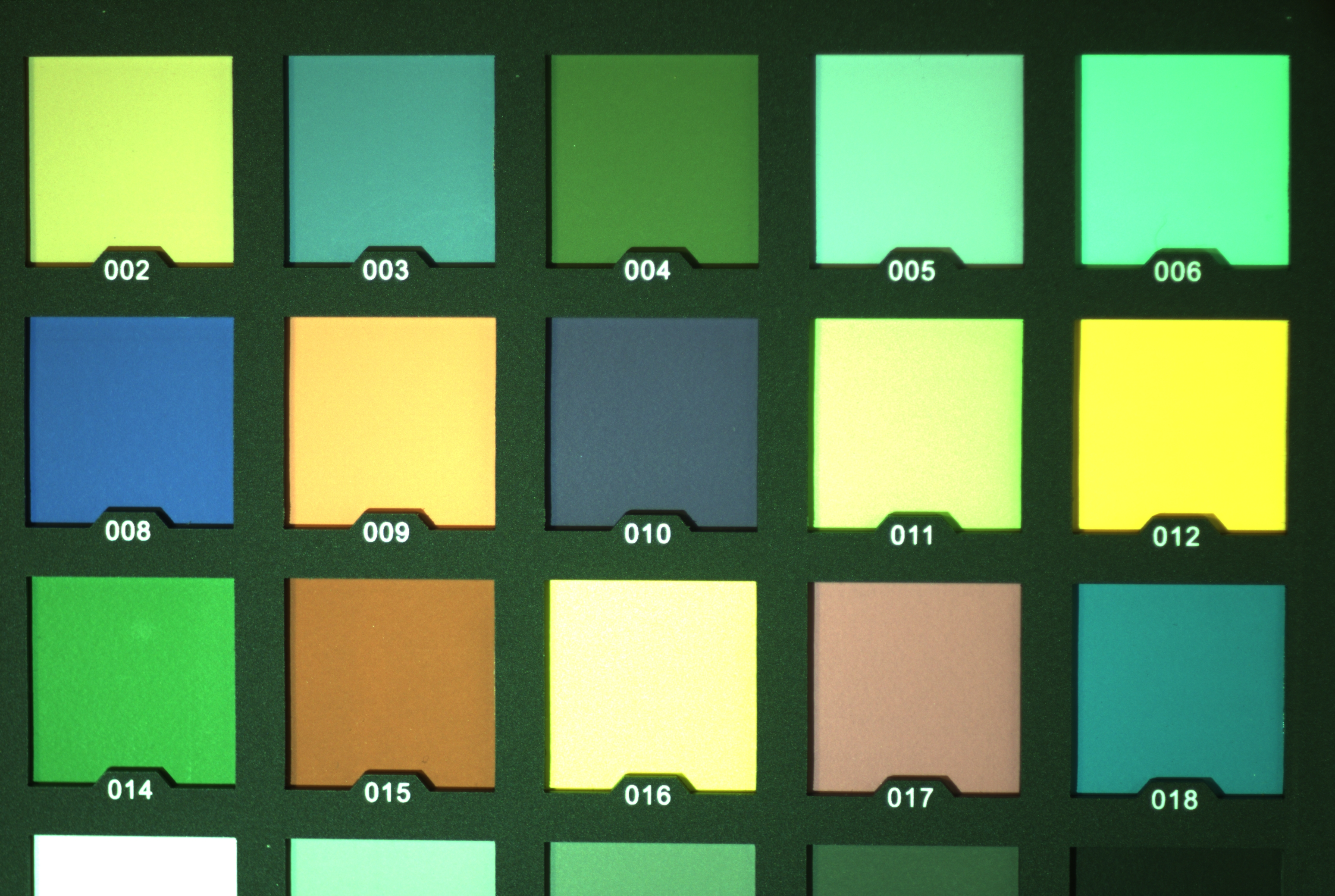
Raw Image
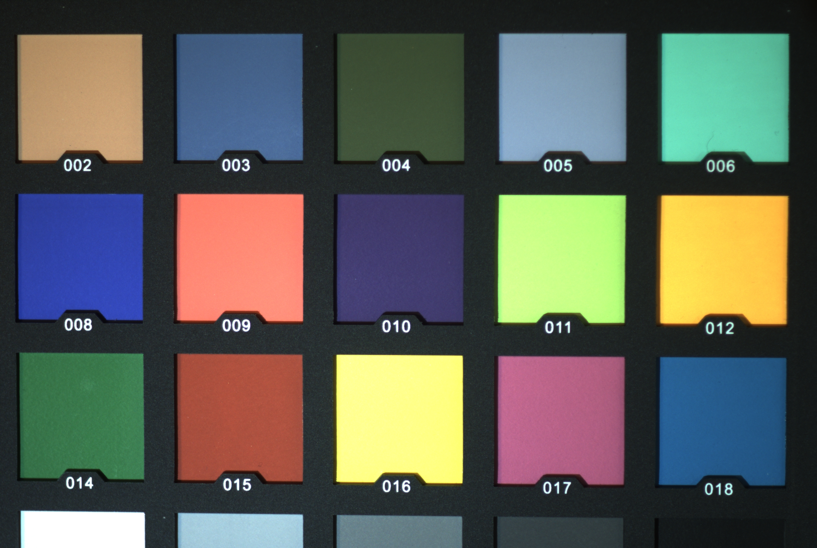
After automatic adjustment
Color adjustment is independent of the camera used.
The algorithm applied works according to our proprietary frugal algorithm approach, which reduces computation time and power consumption.

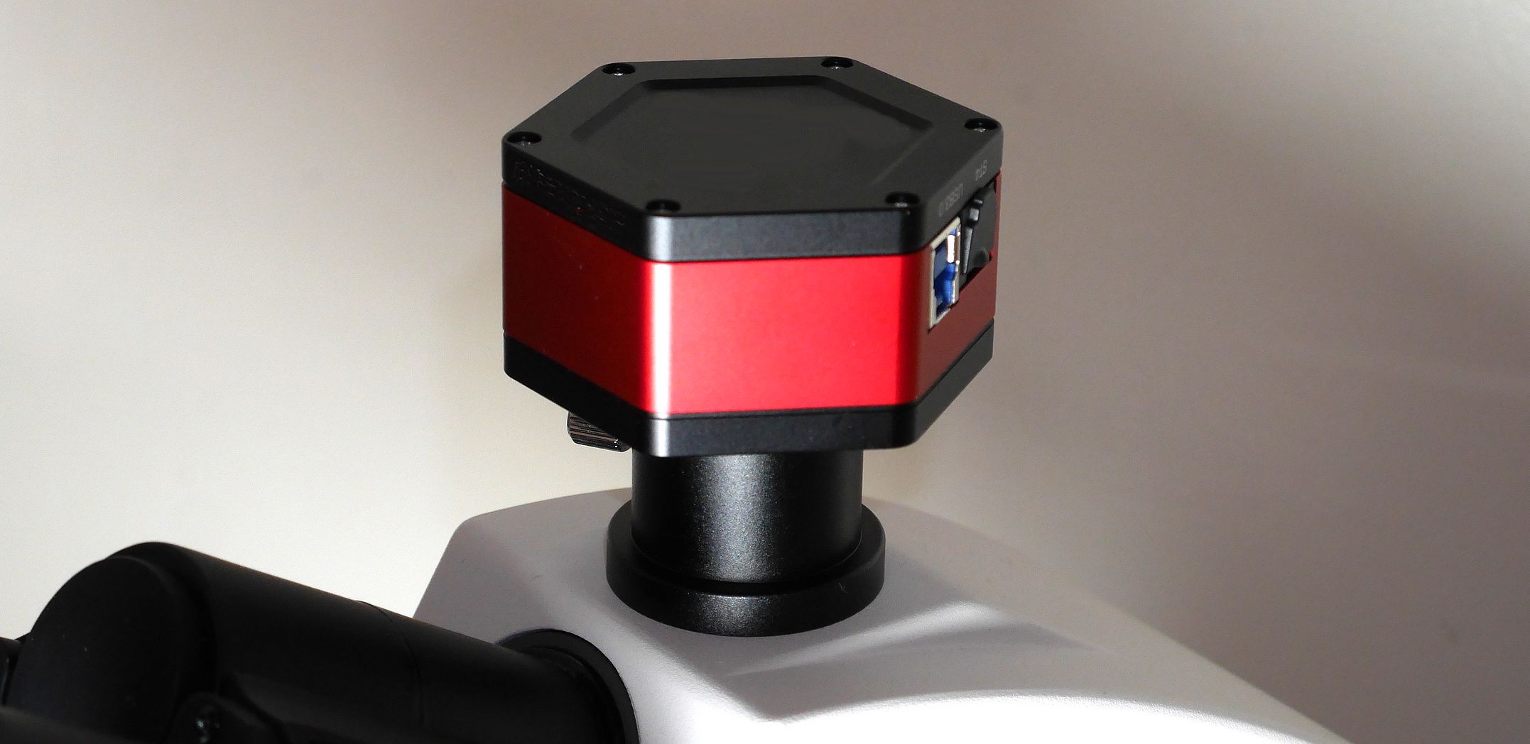
RedCam Cameras
Our cameras have been selected and validated for maximum image quality.
Like all our instruments and components, they are all tested before shipment to ensure their functionality.
Modèle | USB | Taille capteur (mm) | Résolution (pixels) | Taille pixels (µm) | Capteur |
4.2 MPix | 3 | 7.9 x 4.5 | 2,9 | Sony IMX 464 | |
6,3 MPix | 3 | 7,4 x 5,0 | 3096 x 2078 | 2,4 | Sony IMX 178 |
8.3 MPix | 3 | 2,9 | Sony IMX 585 | ||
9.0 MPix | 3 | 3008 x 3008 | 3.79 | Sony IMX 533 |