NEWS
µLight Vision V 1.1.0 is now available!
> New features and tools
> Integration of our RedCam camera
> IDS camera support
> Bug fixes
More information on the “Products / µLight Vision” page
Download the software and user manual from the “Support” page.
Our sources enable structured illumination for transmission microscopy
The µLight technology developed by leida Technologies allows to obtain images impossible to observe with conventional optical microscopy.
The simplicity of structuring the illumination of microscopic preparations in color and shape makes it possible to use it in routine.
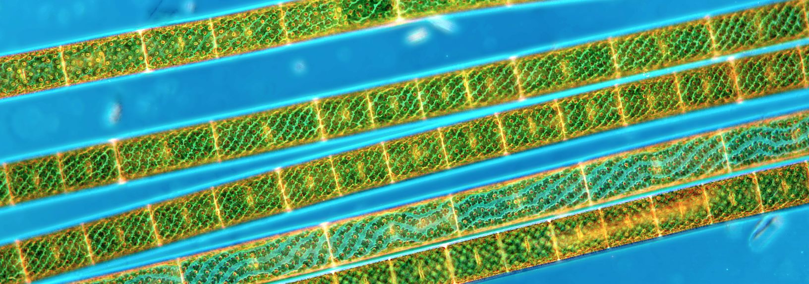
Spyrogira observed in color-coded oblique illumination Courtesy of Martin Gerhardt.
Just click !
The µLight sources are controlled by software. A click on a button allows to modify the lighting structure.
Each image provides different and often complementary information.

Altea pollen grain observed in brightfield, in differential phase contrast and in phase - Courtesy Jean-Pierre Fayol
Multimodalities
The µLight sources allow to observe according to several modalities: brightfield, darkfield, Zernicke phase contrast, differential phase contrast, color coded oblique circular illumination, phase image, Fourier ptychography, digital holography.
A user can easily define his own illumination patterns to develop new methods.
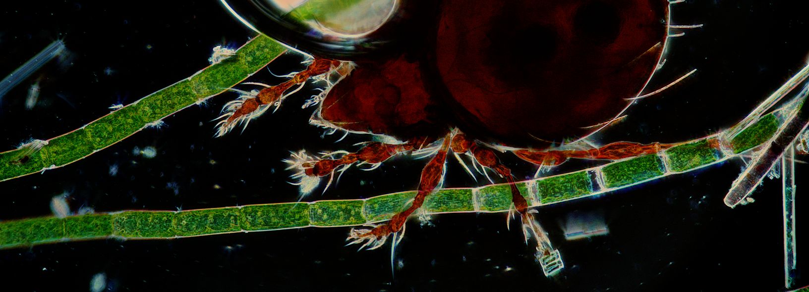
Mite observed against a darkfield- Courtesy of Martin Gerhardt.
Universal
The µLight sources can be installed on almost all microscopes, new and old, upright and inverted.

µLight Gen II - Nikon Eclipse 200
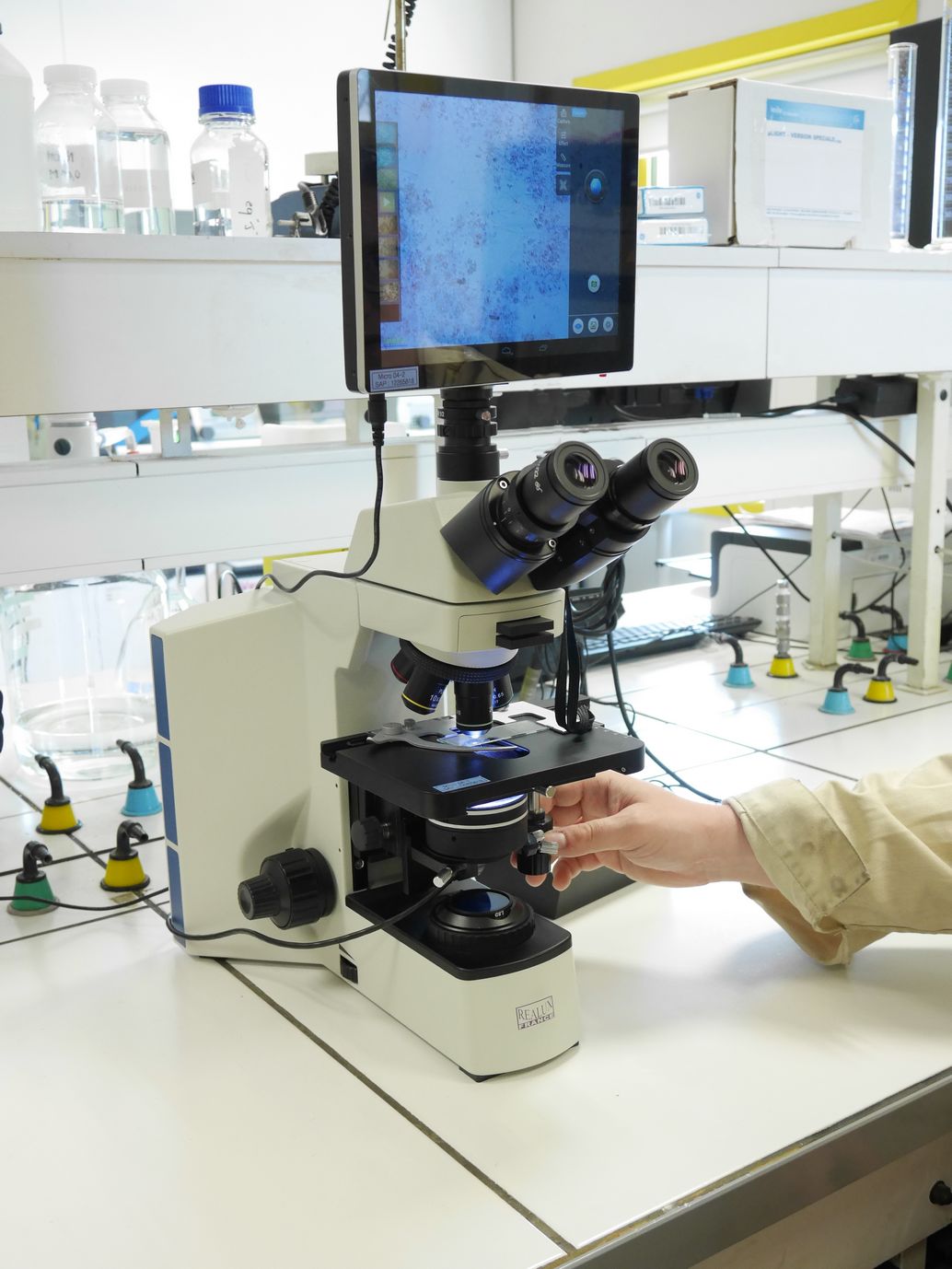
µLigh tLT - CFM-X40 Realux
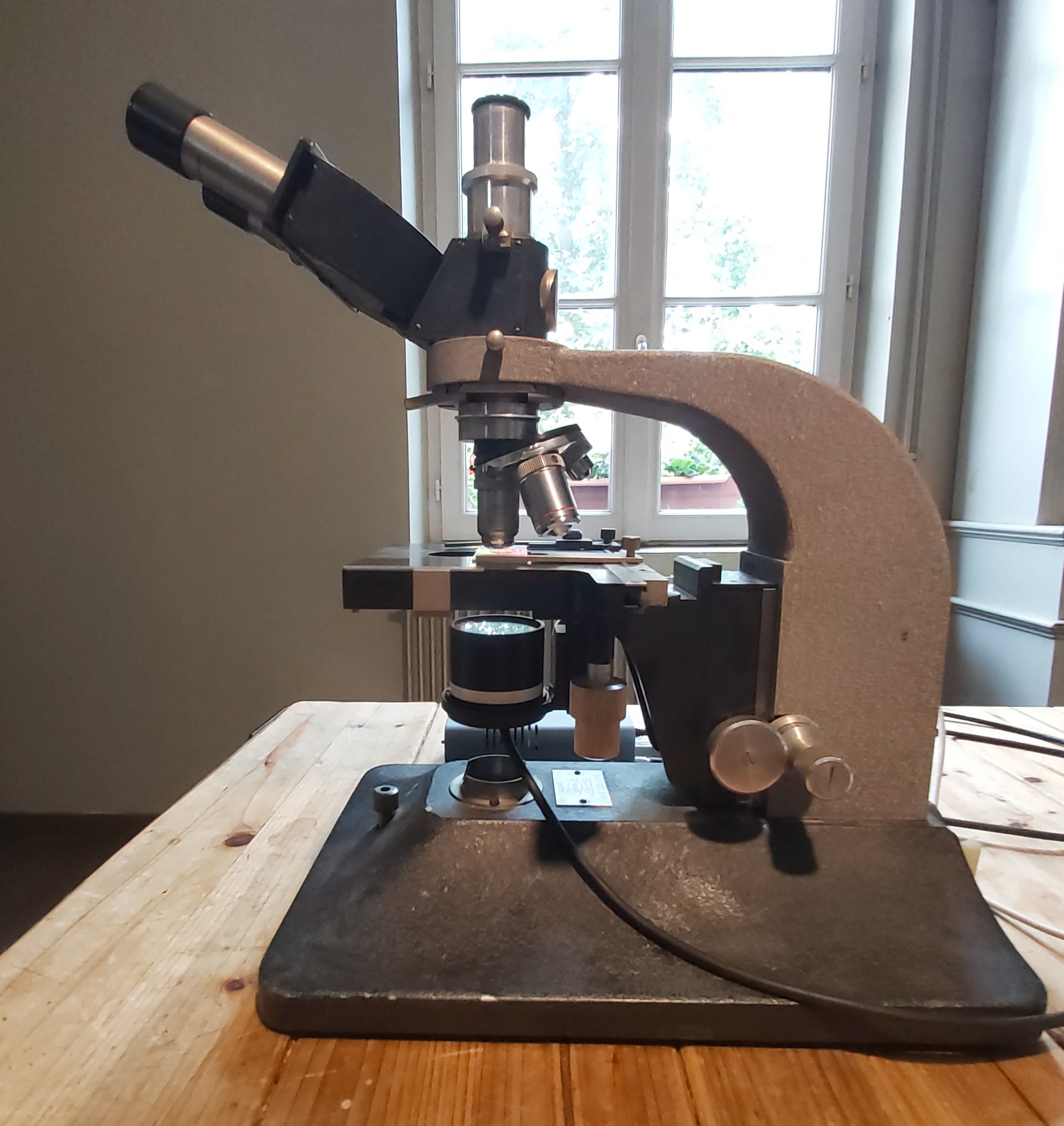
µLight LT2+ - NACHET, very old model

µLight Gen II - Leitz Laborlux

µLight Gen II - BK 5000 Realux
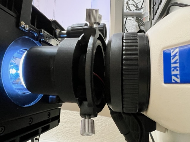
µLight LT2+ - Zeiss Primostar
Our products are distributed exclusively by ARDOP LIGHTING.
Contact: caroline.tovantrang@ardop.com
